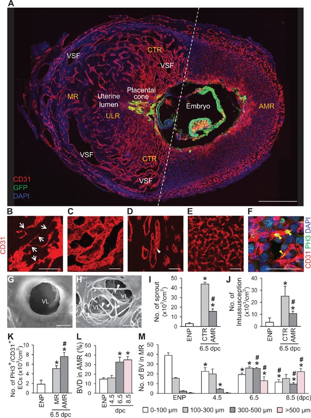
A. Image showing CD31+ blood vessels (BVs) and GFP+ embryo in the uterus at 8.5 dpc. Dotted-line divides the mid-sectioned uterus into the mesometrial region (MR) and anti-mesometrial region (AMR). The MR is subdivided into the central region (CTR) and uterine lumen region (ULR). VSF, vascular sinus folding. Scale bar, 500 µm.
B–F. Images showing CD31+ uterine BVs. Sprouting (B, arrows), variable-sized VSF (C) and intussusceptive pillar (D, white arrowheads) are frequently present in the MR, whereas fine vascular network (E) and PH3+ proliferating ECs (F, yellow arrowheads) are present in the AMR at 6.5 dpc. Scale bars, 50 µm.
G, H. Scanning electron micrographs showing vascular lumens (VL) without and with abundant intussusceptive pillars (arrowheads) in the MR at 6.5 dpc. Scale bars, 5 µm.
I, J. Comparisons of numbers of vascular sprouts and intussusceptions in the uterine endometrium of the estrus stage of non-pregnancy (ENP), and the CTR and AMR at 6.5 dpc. Each group, n = 5–6. *p < 0.002 versus ENP.; #p < 0.001 versus MR by unpaired t test.
K. Comparisons of number of PH3+/CD31+ proliferative ECs in the uterine endometrium of the ENP, and the MR and AMR at 6.5 dpc in a given area (cm2). Each group, n = 5–6 *p < 0.0003 versus ENP; #p = 0.0014 versus MR by unpaired t test.
L, M. Comparisons of CD31+ BV densities (BVD, %) in the AMR and numbers of different sized VSFs in the MR at ENP, and 4.5, 6.5, and 8.5 dpc. Each group, n = 5–6. *p < 0.02 versus ENP.; #p < 0.02 versus 4.5 dpc by one-way ANOVA.