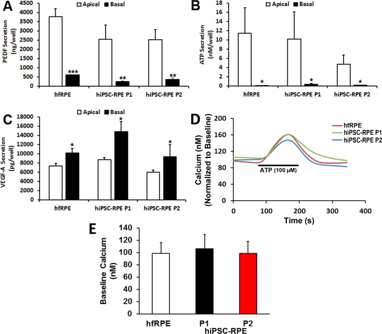
Figure 5.
Polarized factor secretion and G protein–coupled receptor-stimulated calcium responses in passaged hiPSC-RPE. Total amount of (A) PEDF, (B) ATP, and (C) VEGF-A in the apical and basal extracellular media of monolayers of hfRPE and passaged hiPSC-RPE (P1, P2) grown on transwell inserts. (D) Representative traces showing changes in [Ca2+]i in hfRPE and passaged hiPSC-RPE (P1, P2) cultures after stimulation with 100 μM ATP. (E) Baseline [Ca2+]i levels in hfRPE and passaged hiPSC-RPE (P1, P2). *P < 0.05; **P < 0.005; ***P < 0.0005.

