
Figure 1.
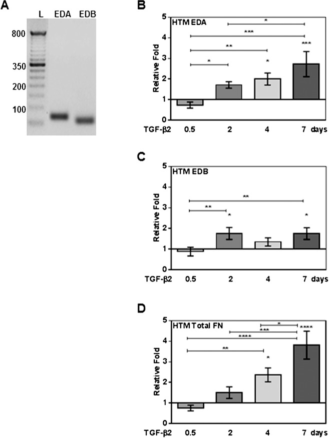
Quantitative RT-PCR analysis of expression, and TGF-β2 induction of cFN and total FN mRNA isoforms in cultured primary human TM cells. Three NTM and four GTM cell strains (HTM cells, n = 7) were treated with or without TGF-β2 (5 ng/mL) for 0.5, 2, 4, and 7 days, and mRNA changes analyzed by qRT-PCR. (A) A representative agarose electrophoresis gel image of EDA and EDB domain qRT-PCR products showing that HTM cells express EDA and EDB cFN isoforms (L, 100 base pair [bp] ladder). Significant induction of EDA (B), EDB (C) isoforms, and total FN (D) mRNA was observed in TGF-β2–treated HTM cells on days 2, 4, and 7. Each FN isoform expression was normalized to β-actin, and mRNA relative fold changes due to treatment were compared to untreated controls (mean ± SEM). Statistical significance between treatment time points were determined using 1-way ANOVA (n = 7; *P < 0.05, **P < 0.01, ***P < 0.001, and ****P < 0.0001).