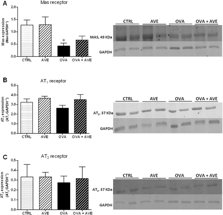
Figure 6.
Expression of angiotensin receptor proteins in lung tissue, using Western blotting (n = 6 in each group). (A) Relative expression of Mas receptor; (B) Relative expression of AT1 receptor. (C) Relative expression of AT2 receptor. After incubation with primary and secondary antibody for Mas, AT1 and AT2 receptors, the membranes were stripped and incubated with primary and secondary antibodies to detect GAPDH; *P < 0.05, significantly different from CTRL or AVE groups; one-way anova followed by Newman–Keuls post test.

