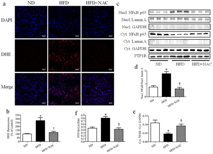
Figure 9. The role of ROS and NFκB in high-fat diet-induced PTP1B expression in skeletal muscle.
Gastrocnemius muscles were isolated from C57 or PTP1BKO mice received normal (ND) or high-fat content (HFD) in a presence or absence of NAC supplementation diets for 3 weeks. (a–b) Representative confocal microscopy pictures (a) and quantification (n = 6) (b) of intracellular ROS production. ROS detection dye DHE is shown as red, the nucleus is stained with DAPI (blue), with purple suggests colocalization. Scale bars, 50 µm. (c–f) Representative Western blots (c) and densitometric analysis (n = 6) of nuclear NFκB (Nucl. NFκB) (d), cytosolic NFκB (Cyt. NFκB) (e), and PTP1B (f) protein expression. * p<0.05 compared with C57 ND mice, † p<0.05 compared with C57 HFD mice.

