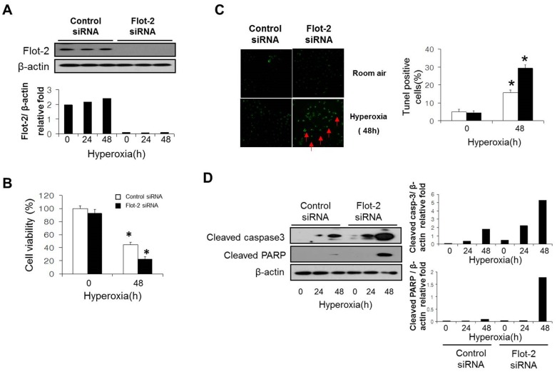
Figure 1. Silencing of Flot-2 promoted cell death and caspase-3 dependent apoptosis.
(A) Beas 2B cells were transfected with Flot-2 siRNA and control siRNA. After 24h, cells were treated with hyperoxia (95% oxygen) or room air. To determine the transfection efficiency, cell lysate was subjected to Western Blot Analysis using anti-Flot-2 antibodies. (B) After another 48h, cell viability was performed using CellTiter-Glo Luminescent Cell Viability Assay kits (Promega). (C) TUNEL staining of Flot-2 silenced Beas 2B cells after hyperoxia. Cells were first transfected with Flot-2 siRNA and control siRNA. As above, cells were exposed to hyperoxia and room air. After 48h, TUNEL staining was performed as described in materials and methods. Left panels: representative images of the TUNEL staining. Apoptotic nuclei were labeled by TUNEL (green, red arrow). Right panel: frequency of TUNEL-positive cells. (D) Deletion of Flot-2 promoted caspase-3 dependent apoptosis. Flot-2 silenced Beas 2B and control cells were exposed to hyperoxia or normoxia (room air) for the indicated time. Cells were harvested and cell lysate was subjected to Western Blot Analysis. Cleaved caspase-3 and cleaved PARP were detected using antibodies specific for the cleaved (active) forms. All the above figures represented three independent repeats with similar results. *P<0.05.

