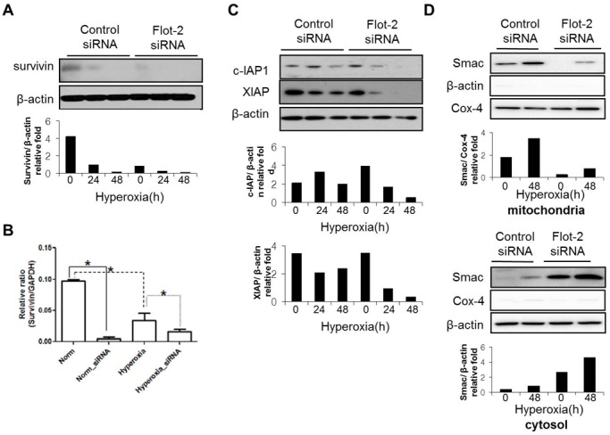
Figure 5. Silencing of Flot-2 down-regulated the expression of IAP family and up-regulated Smac.

Beas 2B cells were transfected with Flot-2 siRNA or control siRNA. Cells were then exposed to hyperoxia or room air. After 24h or 48h, cell lysates were collected for Western Blot Analysis. (A) Protein expression level of survivin. (B) mRNA level of survivin determined by real-time PCR (C) Expression of c-IAP1 and XIAP (D) Silencing of Flot-2 regulated Smac release from mitochondria to cytosol after hyperoxia. As above, Beas 2B cells were transfected with Flot-2 siRNA or control siRNA. After hyperoxia (48h), mitochondria and cytosol were isolated using Mitochondria Isolation Kit as described in material and methods. Equal amount of mitochondria and cytosol were used to perform Western Blot Analysis. Upper panel: Smac detected in mitochondria; Cox-4 was used as the mitochondria marker and β-actin as the cytosol marker. Lower panel: Smac detected in cytosol. All figures above represented three independent experiments with similar results. *P<0.05.
