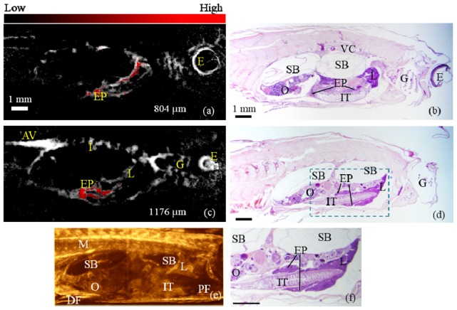
Fig. 6.
(a) & (c): PAT images acquired at the depths of 804 µm and 1176 µm, respectively with the FP labeled exocrine pancreas processed from spectroscopic PAT overlaid in red. (b) & (d): Histological sections at approximate depths as (a) and (c), respectively. (e) is an SD-OCT B-mode image of the zebrafish at approximately corresponding depth to (c). (f) is a zoomed-in display of the dashed region in (d). EP: exocrine pancreas; M: muscle; DF: dorsal fin; PF: pectoral fin; E: eye; AV: aorta and vein; G: gills; I: intersegmental vessels; VC: vertebral column; SB: swim bladder; O: ovary; IT: intestinal tracts; L: liver.

