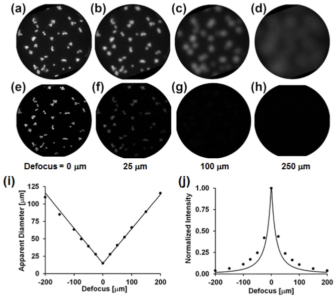
Fig. 2.
HRME (a-d) and confocal microendoscopy (e-h) imaging of a monolayer of 14.8 μm diameter beads as a function of defocus (distance from the monolayer to the fiber bundle’s distal tip). All images are 720 μm in diameter. (i) Measured (dots) and theoretical prediction (line) for bead diameter as a function of defocus. (j) Measured (dots) and theoretical prediction (line) for the mean bead intensity as a function of defocus.

