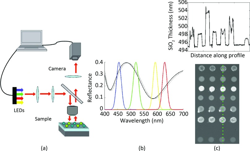
Figure 1.
Summary of the IRIS technique. (a) A schematic of the IRIS platform. LEDs illuminate the sample surface in a reflection mode setup. The surface reflection is imaged to a camera through a 4-f system. (b) Each pixel is recording the spectral intensity information of the surface. The intensity is modulated according to the reflectance corresponding to the SiO2 thickness. (c) Plotting a line profile across a cropped fitted image shows the SiO2 profile of several different proteins.

