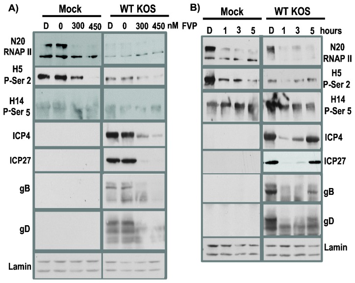
Figure 3. Addition of FVP reduced RNAP II CTD phosphoserine-2 levels and levels of HSV-1 IE proteins ICP4 and ICP27 and late proteins gB and gD were also reduced.
A) HeLa cells were mock infected or infected with WT HSV-1 KOS at MOI of 10. . Flavopiridol (FVP) stocks were prepared in DMSO. At 1 h after infection, DMSO alone or increasing amounts of FVP from 0 to 450 nM were added to cell monolayers. At 8 h after infection, whole cell lysates were prepared and fractionated on 5-15% gradient SDS-polyacrylamide gels. Western blots were probed with N20, H5 and H14 as indicated. Lamin A/C served as the loading control. B) DMSO alone was added at 0 h after infection or FVP (450 nM) was added at 1, 3 or 5 h after infection as indicated. Whole cell lysates were prepared at 8 h. Western blots were probed with anti-RNAP II antibody N20 or phosphoserine-2 antibody H5 or phosphoserine-5 antibody H14. HSV-1 protein ICP4 was detected using antibody P1101; ICP27 was detected with antibody P1119; gB was detected with antibody P1103 and gD was detected with antibody 1123. Lamin A/C served as a loading control.

