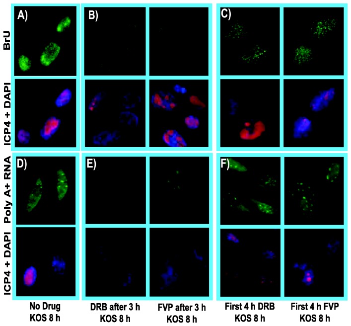
Figure 6. The inhibitory effects of cdk9 inhibiters DRB and FVP on RNA synthesis during HSV-1 infection could be reversed after removing the drugs.
RSF cells were infected with HSV-1 KOS at an MOI of 10. A, D) Infected cells were left untreated. B, E) Infected cells were treated with 100 µM DRB or 450 nM FVP at 3 h after infection. C,F) Infected cells were treated with 100 µM DRB or 450 nM FVP as indicated for the first 4 h of infection after which, cells were washed and drug-free medium was added. A-C). BrU was added to the media at 7.5 h after infection to label nascent RNA. Cells were fixed at 8 h and stained with anti-BrU and anti-ICP4 antibodies and with DAPI. D-F). Cells were fixed at 8 h after infection and in situ hybridization was performed with a biotinylated oligo dT-probe, which was subsequently detected by FITC-conjugated streptavidin. ICP4 staining served as an infection marker and DAPI staining marked nuclei. All images were captured on a Zeiss Axiovert 200M microscope at 100X magnification.

