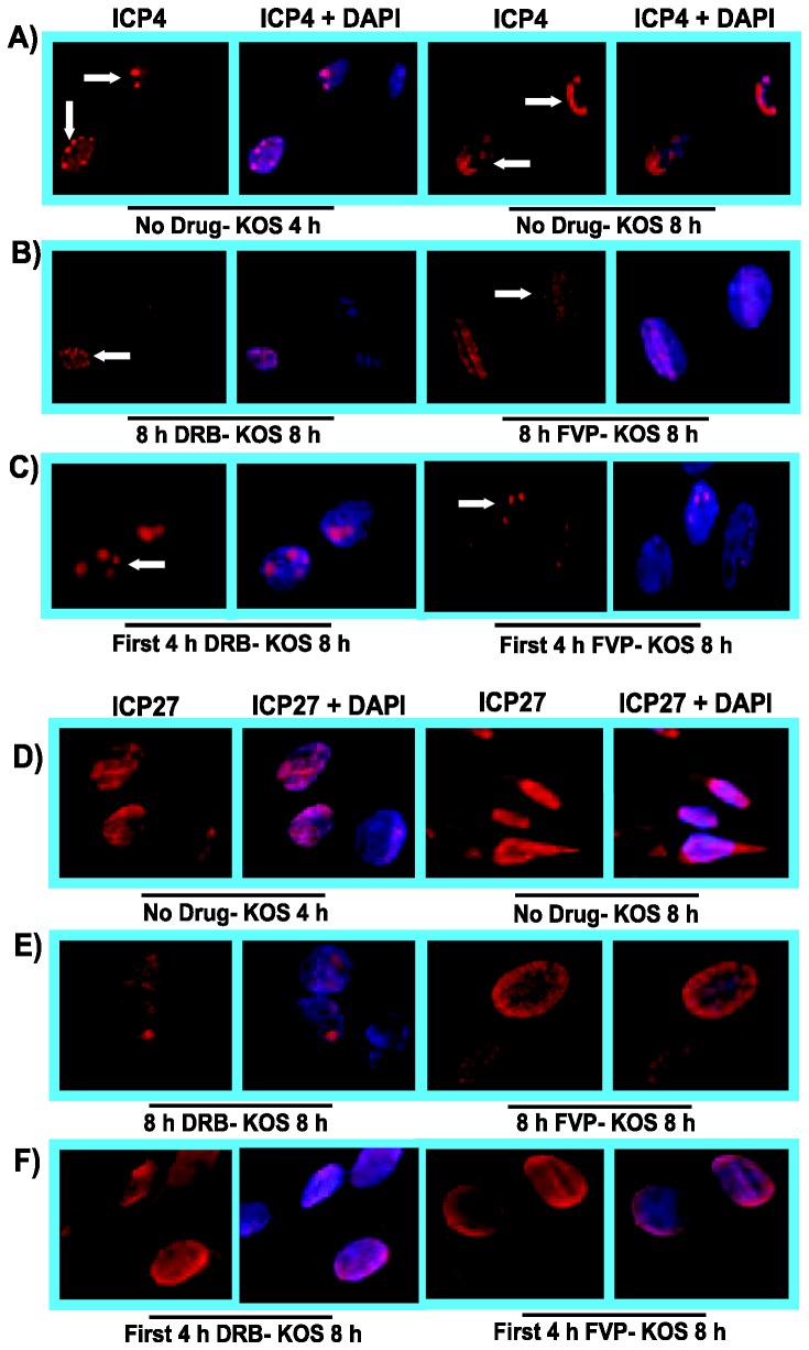
Figure 7. DRB and FVP hindered HSV-1 transcription-replication compartment formation but pre-replicative compartments were apparent after drug removal.
RSF cells were infected with WT KOS at an MOI of 10. A,D) Infected cells were untreated and were fixed at 4 h and 8 h as indicated. B,E) Cells were treated with 100 µM DRB or 450 nM FVP as indicated at the beginning of infection and were fixed at 8 h. C,F) DRB or FVP were added at the start of infection and were removed at 4 h and cells were incubated in drug-free medium for an additional 4 h. Infected cells were fixed at 8 h. A,B,C). Cells were stained with anti-ICP4 antibody to monitor viral transcription-replication compartment formation and DAPI to mark nuclei. D,E,F). Infected cells were stained with anti-ICP27 antibody to monitor ICP27 sub-cellular localization and DAPI to mark nuclei. Images were captured on a Zeiss Axiovert 200M microscope at 100X magnification.

