
Figure 9. Viral replication was greatly reduced in the presence of DRB and FVP but viral replication resumed when the drugs were removed after 4 h.
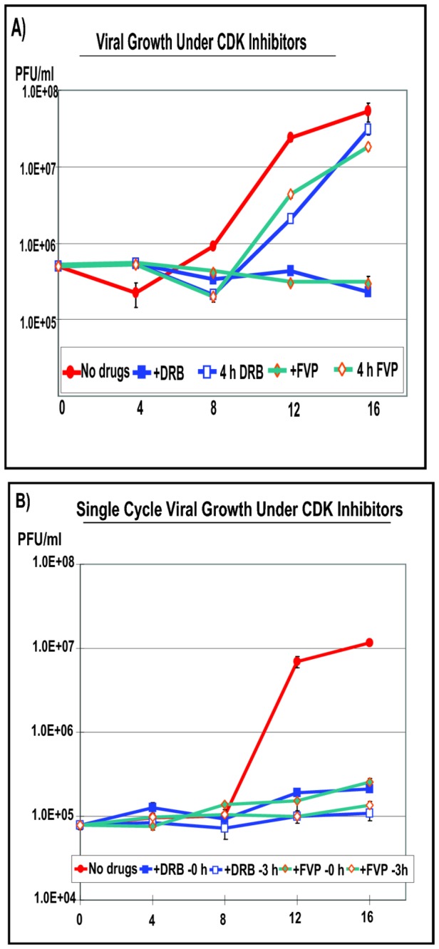
HeLa cells were infected with HSV-1 KOS at MOI of 1. A) Infected cells were either untreated (no drugs) or DRB (100 µM) or FVP (450 nM) were added at the time of infection for the duration of infection, or DRB and FVP were present for the first 4 h and then were removed and cells were incubated in drug-free medium for the remainder of the time, as indicated. Samples were harvested at 0, 4, 8, 12 and 16 h after infection and virus titers were determined by plaque assays. B) HeLa cells were infected with KOS at an MOI of 1 and were untreated (no drugs) or treated with DRB or FVP starting at 0 h or 3 h as indicated and the drugs were present for the duration of the experiment. Samples were harvested at 0, 4, 8, 12 and 16 h as described in panel A. The experiments were performed in triplicate and error bars are shown.