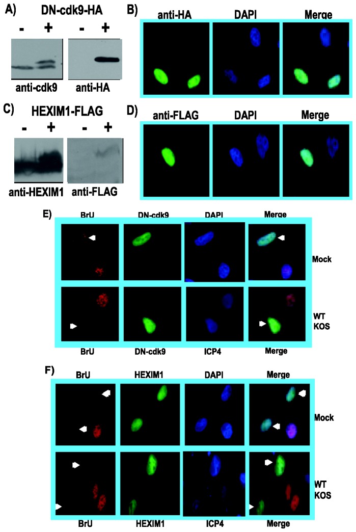
Figure 10. Expression of a dominant-negative kinase-dead cdk9 mutant or the cdk9 negative-regulator HEXIM1 inhibited nascent RNA synthesis.
A) HeLa cells were either transfected with empty vector or with 2 µg of DN-cdk9-HA DNA, which expresses an HA-tagged kinase-dead cdk9 dominant negative mutant. After 24 h, whole cell lysates were prepared and fractionated by SDS-PAGE. Western blots were probed with anti-cdk9 antibody (left panel) or anti-HA antibody (right panel). B) HeLa cells transfected with DN-cdk9-HA were fixed 24 h after transfection and stained with anti-HA antibody to visualize DN-cdk9-HA and DAPI to mark nuclei. C) HeLa cells were transfected with empty vector or with plasmid DNA expressing FLAG-tagged HEXIM1. Whole cell lysates were prepared as in panel A and Western blots were probed with anti-HEXIM1 antibody (left) and anti-FLAG (right). D) Indirect immunofluorescence was performed on HeLa cells transfected with HEXIM1-FLAG. Anti-FLAG antibody was used to visualize HEXIM1-FLAG and DAPI staining marked the nuclei. E) HeLa cells were transfected with DN-cdk9-HA for 24 h and were subsequently mock infected or infected with HSV-1 KOS. Bromouridine (BrU) was added at 7.5 h after infection for 30 min, at which time cells were fixed and stained with anti-BrU antibody to visualize newly transcribed RNA and anti-HA antibody to visualize DN-cdk9-HA. DAPI staining in the upper mock panels marked nuclei and staining with anti-ICP4 antibody in the lower KOS panels was used as a marker for infection. White arrows point to cells expressing DN-cdk9-HA in the BrU and merged panels. F) HeLa cells transfected with HEXIM1-FLAG for 24 h were mock infected or infected with HSV-1 KOS and BrU was added at 7.5 h after infection for 30 min. Cells were fixed at 8 h and stained with anti-BrU to detect newly transcribed RNA and anti-FLAG antibody to detect HEXIM1-FLAG. DAPI was used to mark nuclei in the upper mock panels and anti-ICP4 antibody was used in the lower KOS panels as a marker for infection. White arrows point to cells expressing HEXIM1-FLAG. All images were captured on a Zeiss Axiovert 200M microscope under 100X magnification.

