
Figure 1. SICA protein profile from Pk1(B+)1+ and Pk1(B-)1- immunoprecipitations.
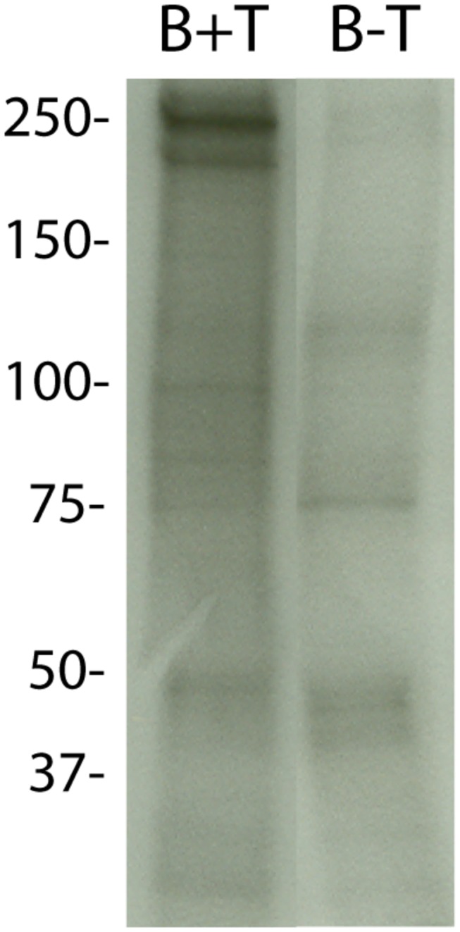
Trophozoite SDS extracts were immunoprecipitated with purified IgG from an antiserum (Cyto17) (Al-Khedery, 1999) that is specific for the SICA conserved cytoplasmic domain. The intense bands visible around 250 kD in the Pk1(B+)1+ parasites but lacking in the Pk1(B-)1- clone represent the SICA proteins.