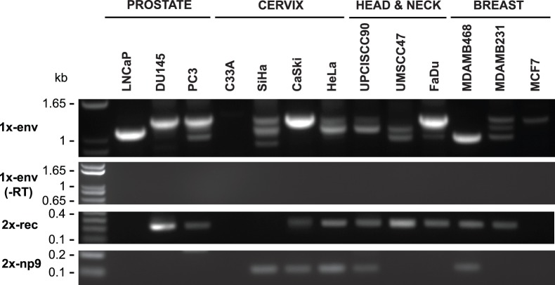
Figure 3. Detection of HERV-K transcripts in 13 cancer cell lines.
Nested PCR was performed on RT-PCR products shown in Fig. 2 to detect viral transcripts at three different positions in HERV-K genome. Two of RT-PCRs cross splicing junctions to detect RNAs spliced at the conventional env mRNA splice junction (1x-env) and rec and np9 mRNA (2X-rec, 2X-np9) splice junctions. The products were resolved by electrophoresis. Genomic positions of the primers used are shown in Fig. 1. Parallel controls were performed without reverse transcriptase (-RT) as controls to exclude DNA contamination. DNA size markers are shown on the left.

