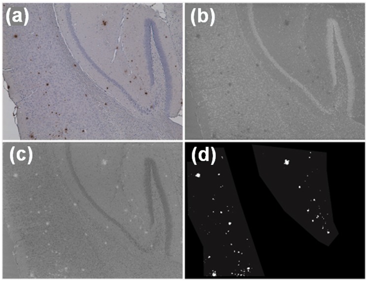
Figure 6. Visualising brain plaques using image segmentation methodology with Aβ42-specific 1E8 antibody.
Original RGB image of typical transgenic brain section showing significant plaque presence (a) and nuclear image obtained using colour unmixing; plaques are mostly absent from this image (b). Plaque image obtained by colour unmixing; nuclei are mostly absent from this image while plaques are clearly visible (c). Masks for the cortex area and the hippocampus were produced manually as shown by lighter grey shading (d). Plaques shown in white were segmented automatically from image (c) using intensity Otsu thresholding on image (b). All images are 20×magnification.

