
Figure 7. GW843682X (GW) down-regulates TNF-α expression via inhibition of MAPK and NF-κB signaling.
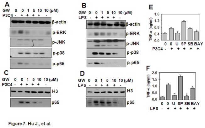
(A, B) The effect of GW on Pam3CSK4- and LPS-induced signal transduction. THP-1 cells, starved overnight and pre-treated with the indicated concentrations of GW for 30 min, were re-stimulated with 1 µg/ml Pam3CSK4 (P3C4) (A) or LPS (B) for 30 min. The phosphorylation of ERK, JNK, p38, and NF-κB p65 was detected by western blot. β-actin protein expression was detected as loading control. (C, D) The effect of GW on Pam3CSK4- and LPS-induced NF-κB p65 nuclear localization. THP-1 cells were treated as in (A) (C) and (B) (D). Total proteins in the nucleus were extracted, and NF-κB p65 protein was detected by western blot. Histone H3 protein expression was detected as a loading control. (E, F) Pam3CSK4 and LPS induced TNF-α via the MAPK and NF-κB pathways. THP-1 cells, pre-treated with 10 µM U0126 (U), or SP600125 (SP), or SB203580 (SB), or Bay11-3072 (BAY) for 30 min, were re-stimulated with 1 µg/ml Pam3CSK4 (P3C4) (E) or LPS (F) for 24 h. TNF-α secreted in the supernatant was detected by ELISA. * P < 0.05 compared with the Pam3CSK4- or LPS-treated groups.