
Figure 2. CyPPA prevents cytokine release.
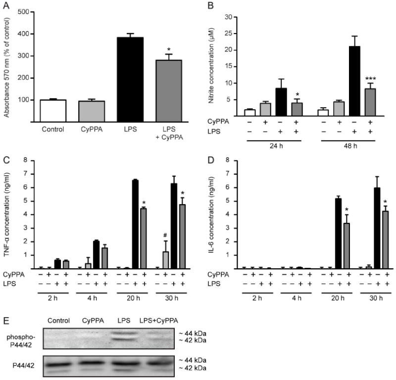
A. MTT analysis of microglial cells treated with CyPPA (25 μM) for 24 in the presence of LPS (200 ng/ml). Results shown represent mean ± S.D. (*p<0.05 versus LPS-treated microglia, ANOVA, Scheffé’s test, n=6 wells, experiment repeated at least 3 times with independent primary microglia preparations). B. NO production of microglial cells treated with 200 ng/ml LPS in the presence and absence of CyPPA (25 μM). The effects of CyPPA (25 μM) on cytokine production, TNF-α (C) and IL-6 (D) in LPS (200 ng/ml)-activated microglia for 2-30 h. Results represent mean S.D. (*p <0.05; ***p<0.001 versus LPS-treated microglia; #p<0.05 versus non-treated cells, U-test Mann-Whitney, n=3). E. Western blot analysis of phosphorylated and non-phosphorylated p44/p42 MAPK in microglial cells treated in the presence or absence of CyPPA (25 μM) and LPS (200 ng/ml).