
Figure 6. KCa2.3 regulates microglial activation pathways.
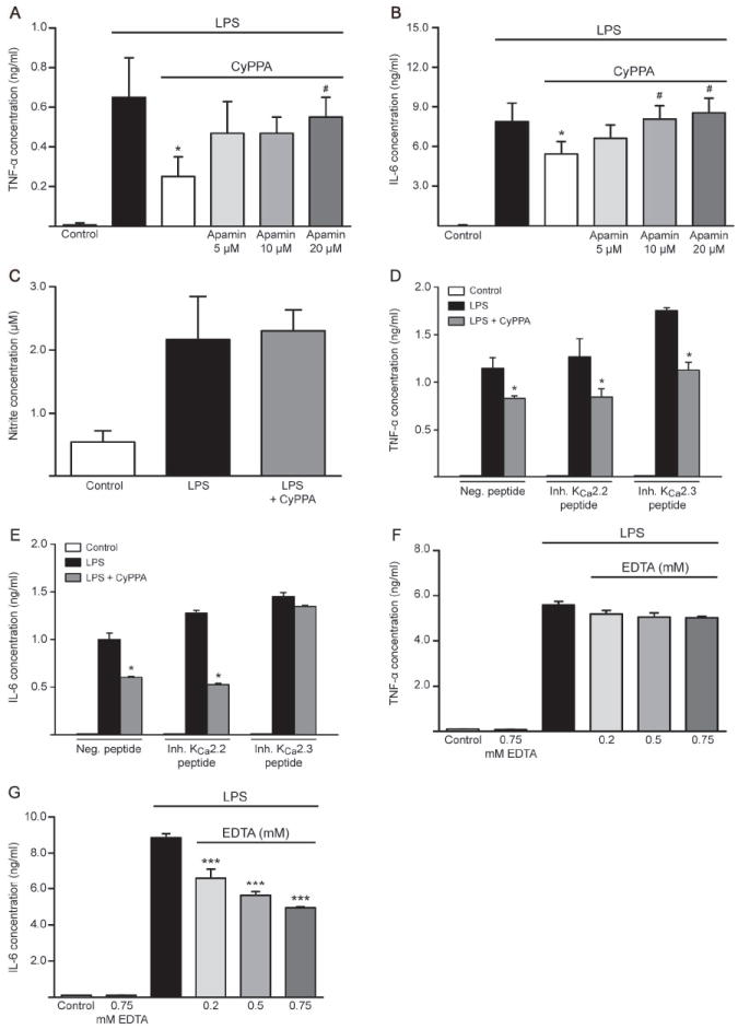
Cytokine production, TNF-α (A) and IL-6 (B) in microglial cells co-treated with LPS (200 ng/ml) and CyPPA (25 μM) in the presence and absence of different concentrations of apamin. Results represent mean ± S.D. (*p <0.05 versus LPS-treated microglia, #p<0.05 versus CyPPA-treated microglia, U-test Mann-Whitney, n=3, experiment repeated 3 times with independent primary microglia cultures). C. NO release in microglial cells transfected with inhibitory peptides for KCNN3/SK3/KCa2.3 channels. Results shown represent mean ± S.D. (n=3, experiment repeated 3 times with independent primary microglia cultures). D. TNF-α and E. IL-6 production in microglial cells transfected with inhibitory peptides for KCNN2/SK2/KCa2.2 and KCNN3/SK3/KCa2.3 channels and challenged with LPS (200 ng/ml) in the presence of CyPPA (25 μM). Results are presented as mean values ± S.D. (*p <0.05 versus LPS-treated microglia, U-test Mann-Whitney, n=3, experiment repeated 3 times with independent primary microglia cultures). F. TNF-α and G. IL-6 production in microglial cells and co-treated with different concentrations of EDTA, as indicated. Results are shown as mean values ± S.D. (***p <0.001 versus LPS-treated microglia, U-test Mann-Whitney, n=6, experiment repeated 3 times with independent primary microglia cultures).