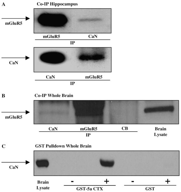
Fig. 6.
(A) Coimmunoprecipitation of mGluR5 and calcineurin. Immunoblots of rat hippocampal homogenates were immunoprecipitated with either mGluR5 or calcineurin (CaN) antibodies, and probed with mGluR5 and CaN antibodies. The blots show that mGluR5 and CaN are immunoprecipitated from hippocampal lysates, and that CaN coimmunoprecipitates with mGluR5, and mGluR5 coimmunoprecipitates with CaN. (B) Immunoblot of rat whole brain lysates immunoprecipitated with CaN, mGluR5, or a negative control cascade blue antibody, and probed for mGluR5. The blot shows that mGluR5 is detected in total brain lysate and in the mGluR5 immunoprecipitate, and that CaN is also detected as a coimmunoprecipitate of mGluR5, however, there is no mGluR5 detected in the negative control (CB). (C) Pull-down assay of mGluR5a and CaN. Rat whole brain homogenates were incubated with GST fusion protein or GST-mGluR5a carboxy terminal domain (GST-5aCTX) and probed with CaN antibody. The results show no CaN detected in GST alone, GST incubated with rat brain lysate, or GST-5aCTX alone, but CaN is detected in total brain lysate and GST-5aCTX incubated with rat brain lysate.

