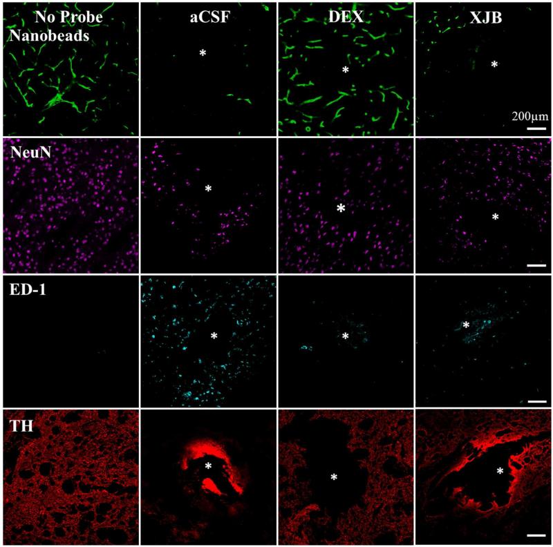
Figure 4.
DEX and XJB mitigate the histochemical impact of penetration injury. Separate columns provide representative images of tissue after retrodialysis of aCSF, DEX, and XJB for 4 h. The left-most column shows images of non-implanted control striatal tissue. Separate rows provide representative images of tissue labeled with markers for blood flow (nanobeads), neuronal nuclei (NeuN), macrophages (ED-1), and dopamine axons and terminals (TH). The probe track is in the center of the images and marked with an asterisk. Scale bars are 200 μm.

