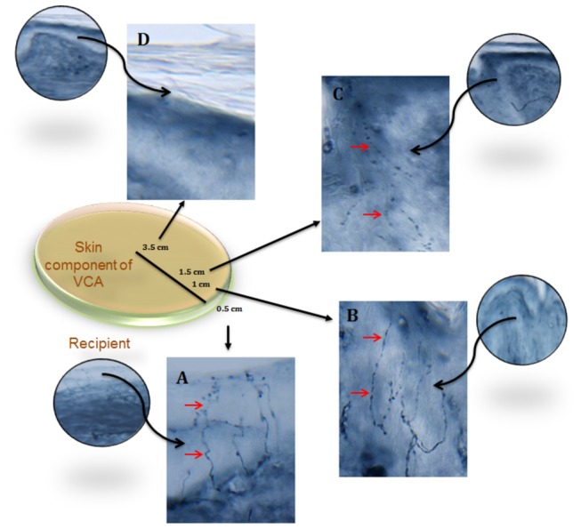
Figure 5. Sequential sections of skin component of allograft (Day 240 post-transplant).

PGP9.5 (pan-axonal marker) demonstrates dense epidermal fibers (solid arrow) in native skin 0.5 cm away from the graft (A). Intra-epidermal fibers also visualized in skin component of VCA at 1cm (B) and 1.5 cm (C) away from the junction with native skin. No epidermal fibers seen in the center of alloflap (D).
