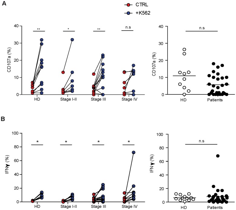
Figure 2. Ex vivo NK cell functionality in donors and melanoma patients.
Patients were stratified (left panels) or not (right panels) according to their clinical stage. Freshly isolated PBMC were stimulated with K562 (E/T ratio 10/1 for 4 hours) in absence of cytokine and gated CD3−CD56+ NK cells were analyzed by flow cytometry. (A) Percentages of NK cell degranulation and (B) IFNγ production were assessed. On left panels, spontaneous (red) and K562-activated (blue) values are indicated. On right panels, the effective NK cell activation was reported calculated as K562-induced minus spontaneous. Comparisons between spontaneous (ctrl) and K562 induced activation of NK cells were analyzed with the paired t-test (p values≤0.05 noted as * and p<0.01 as **). Comparison of NK function between donors and patients (right panels) using the non parametric Mann-Withney test (n.s = not significant).

