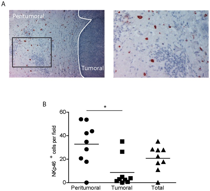
Figure 6. NK cells are present in primary skin melanoma microenvironment.
The presence of NK cells (in red) was analyzed in paraffin-embedded thin melanoma sections by anti-NKp46 immunohistochemical labeling. (A) One representative image is reported. Original magnifications: ×10 (left panel) and ×20 (right panel) followed by computer magnification. (B) NKp46+ cells were counted in 10 tumoral and 10 peritumoral fields as described in material and methods. Results are expressed as mean values. Horizontal lines represent median values. NK cell numbers were compared with paired t test (p = 0.048).

