
Fig. 4.
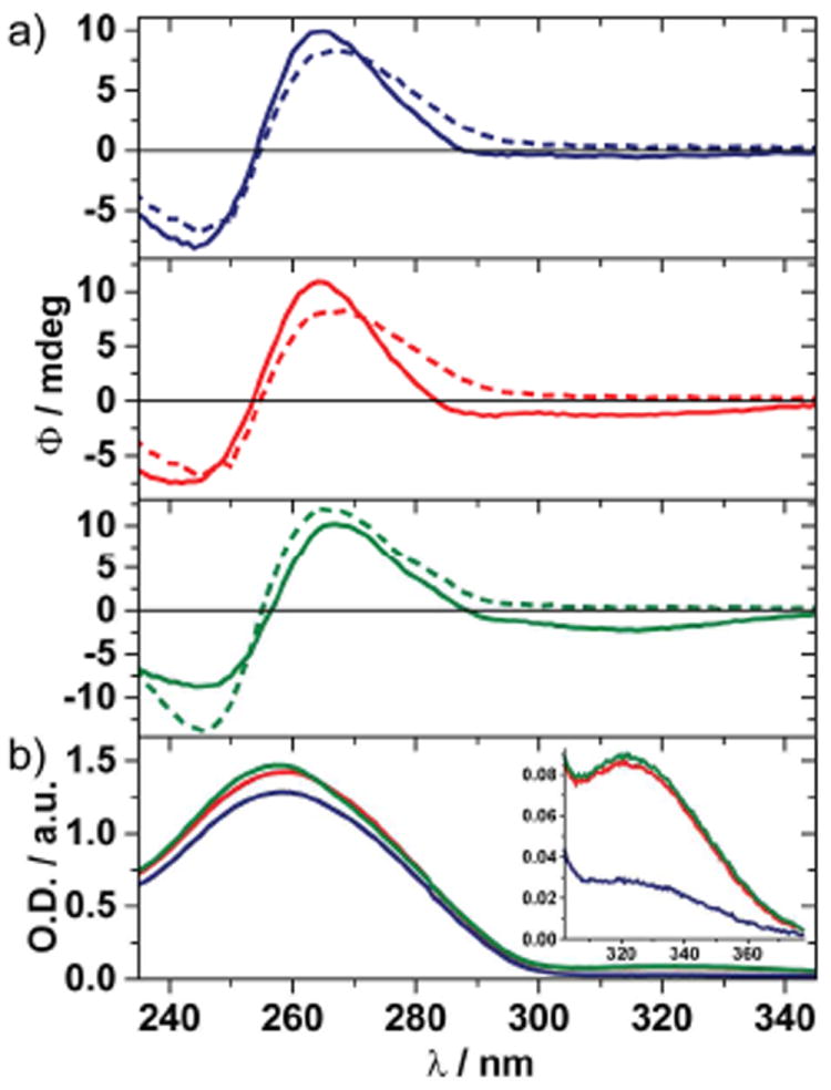
a) CD spectra, and b) absorption spectra of 3•6 (blue), 4•6 (red), 5•7 (green), and CD spectra of native control duplexes (dashed lines). Samples consist of 25 μM dsDNA in 10 mM phosphate buffer containing 100 mM NaCl at pH 7. Fluorimeter settings: λex = 332 nm and 5 nm excitation and emission slit widths.