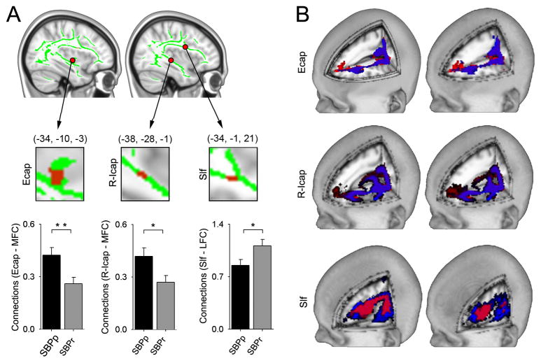
Figure 4. Differential structural connectivity to medial and lateral prefrontal cortex for tracts where FA was lower in SBPp.
(A) White matter seeds (Ecap, R-Icap, Slf, red; green = white matter skeleton; derived from regions with decreased FA in SBPp in discovery-SBP at visit 1), that differentially connect with medial (MFC) and lateral (LFC) prefrontal cortex (tested for all SBP, n=46). In SBPp, Ecap (t44 = 2.87, p = 0.006) and r-lcap (t44 = 2.38, p = 0.022) show higher connectivity to the medial prefrontal cortex; while in SBPr, slf shows higher connectivity to lateral prefrontal cortex (t44 = −2.223, p = 0.031). (B) Tractography schematic showing group mean tracts seeded from Ecap and r-Icap more connected to MFC in SBPp (red) whereas the group mean tracts seeded from slf are more connected to the LFC in SBPr (blue). *P < 0.05, **P < 0.01. Error bars represent S.E.M. slf= superior longitudinal fasciculus; Ecap =external capsule; r-Icap= retro-lenticular limb of internal capsule.

