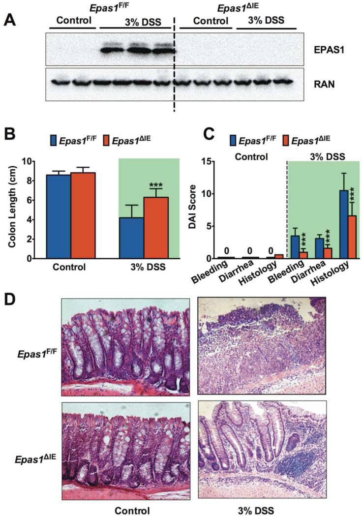
Figure 2. Disruption of intestinal EPAS1 protects mice from DSS-induced colitis.
(A) Western blot analysis of EPAS1 in colons from Epas1F/F and Epas1ΔIE mice treated with 3% DSS or regular water (Control) for 3-days. (B) Colon lengths, (C) disease activity index (DAI) and (D) H&E staining of colon tissue after 3% DSS or Control treatment for 7 days in Epas1F/F (n=15) and Epas1ΔIE (n=16) mice. Shaded areas highlight the 3% DSS treatment. Each bar represents the mean value ± S.D.***p<0.001, compared to Epas1F/F mice.

