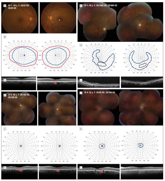
Figure 2.
Clinical features in the affected family members. A, D, G, and J, Color fundus photographs. B, E, H, and K, Goldmann perimetry. C, F, I, and L, Spectral-domain optical coherence tomographic images through fixation, except in family member IV-1, where black lines indicate vertical scan location. Homozygotes show outer retinal loss with hyperreflective lesions in the outer nuclear layer (black arrows). The asterisk indicates a region of mild retinal pigment epithelial (RPE) depigmentation; red arrows, disrupted reflectivity of the outer segment–RPE junction and hyperreflective lesions external to the external limiting membrane and inner-segment ellipsoid band. OD indicates right eye; OS, left eye.

