
Figure 5. Expression profile of Hbl proteins in B. cereus ATCC 10876.
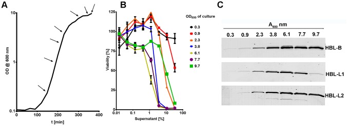
(A) Growth measurements (OD at 600 nm) of B. cereus ATCC 10876 grown in Brain Heart Infusion broth over time. Arrows indicate points at which samples were withdrawn for analyses performed in (B) and (C). (B) Viability assessment of macrophages incubated for 90 min with B. cereus ATCC 10876 sterile, serially-diluted culture supernatants. (C) Western blot analysis of growth-phase dependent expression and accumulation of Hbl toxin components B, L1, and L2 of B. cereus ATCC 10876 in sterile culture supernatants. Toxin proteins were detected using mouse serum containing antibodies raised against single toxin components.