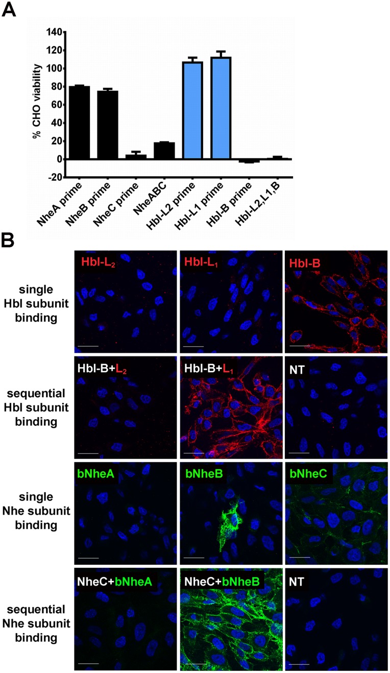
Figure 7. Sequential binding of toxin components to cells.
(A) Viability of CHO cells primed for 15 min with either one of the three Nhe (black bars) or Hbl (blue bars) toxin components (5 nM for Hbl toxins, 3% final concentration of recombinant Nhe toxin-containing sterile culture supernatants), followed by washes and challenge with the two complementary components for 30 min (“prime”), or treated with a mixture of all three proteins (A,B,C or L2,L1,B). Viability is shown relative to control wells treated with the two complementary challenge components only. (B) Confocal microscopic evaluation of toxin component binding to CHO cells. DAPI-stained nuclei are shown in blue. Cell-bound Hbl toxin subunits were detected using antibodies raised against the specific subunit, followed by a secondary, Alexa-Fluor 594 labeled antibody (upper two rows). The specific protein detected is indicated by a red label. Binding of (biotinylated, indicated by a “b” in the toxin name) Nhe toxin subunits was detected by using streptavidin labeled with Alexa Fluor 488 (lower two rows). The detected protein is shown is indicated by a green label. NT samples represent cells not treated with toxin, incubated with the Alexa Fluor labeled antibody alone. Bar represents 20 µm.

