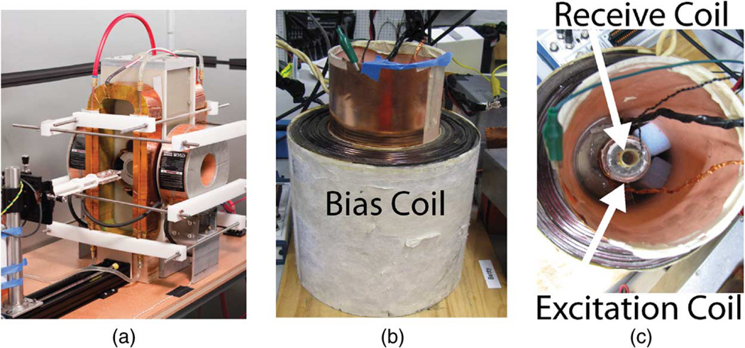
Fig. 2.
Berkeley x-space projection MPI scanner (a) acquires 2-D images. A 2.3 T/m magnetic gradient creates a FFL, and the excitation coil scans this FFL at 22.9 kHz with field strengths up to 35 mT-pp. The Berkeley x-space relaxometer, shown with side (b) and top (c) views, measures the point spread function of a particle sample. The excitation coil generates a sinusoidal magnetic field of 10–200 mT-pp strength at frequencies of 1.5–11.5 kHz. The signal received from the gradiometric receive coil is digitized at 10 MSPS without filtering. The bias coil can add ± 180 mT field for partial FOV scanning.

