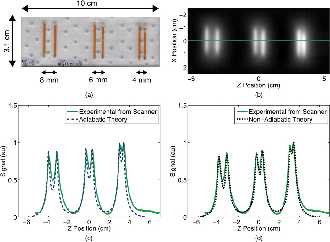
Fig. 4.
A line resolution phantom (a) was constructed with 1.75-mm-wide wells which were filled with 20 × diluted Resovist. We acquired a positive-velocity scan image (b) of this phantom in the Berkeley x-space projection MPI scanner at 20 mT-pp for a scan time of 59 s and a FOV of 13 cm 5 cm. No deconvolution was performed. We visualized a 1-D profile through the center of this image and compared this profile to the image predicted by the adiabatic x-space theory (c) and by the non-adiabatic x-space theory (d). The experimentally measured image showed better agreement with the non-adiabatic x-space theory than with the adiabatic x-space theory. Non-adiabatic calculations used a measured relaxation time constant of 2.9 µs.

