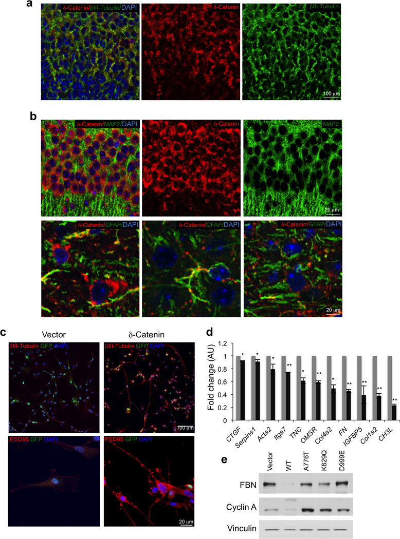
Figure 4.

Expression of δ-catenin in neurons and δ-catenin driven loss of mesenchymal marker in GBM. a, Pattern of expression of δ-catenin in the developing brain, as determined by immunostaining. Double immunofluorescence staining of brain cortex using δ-catenin antibody (red) and βIII-tubulin (green); Nuclei are counterstained with Dapi (blue). b, Pattern of expression of δ-catenin in the adult brain, as determined by immunostaining. Upper panels, Double immunofluorescence staining of brain cortex using δ-catenin antibody (red) and MAP2 (green); Nuclei are counterstained with Dapi (blue). Lower panels, Double immunofluorescence staining of of brain cortex using δ-catenin antibody (red) and GFAP (green); Nuclei are counterstained with Dapi (blue). c, Immunofluorescence staining for βIII-tubulin (upper panels) and PSD95 (lower panels) in U87 cells expressing δ-catenin or the empty vector. d, Expression of mesenchymal genes in glioma cells expressing δ-catenin or the empty vector (averages of triplicate quantitative RT-PCR). Error bars are SD p is from t-test. *, p ≤ 0.005; **, p ≤ 0.001. e, Western blot using the indicated antibodies for U87 cells expressing δ-catenin wild type, glioma–associated δ-catenin mutants or the empty vector. FBN, fibronectin. Vinculin is shown as control for loading.
