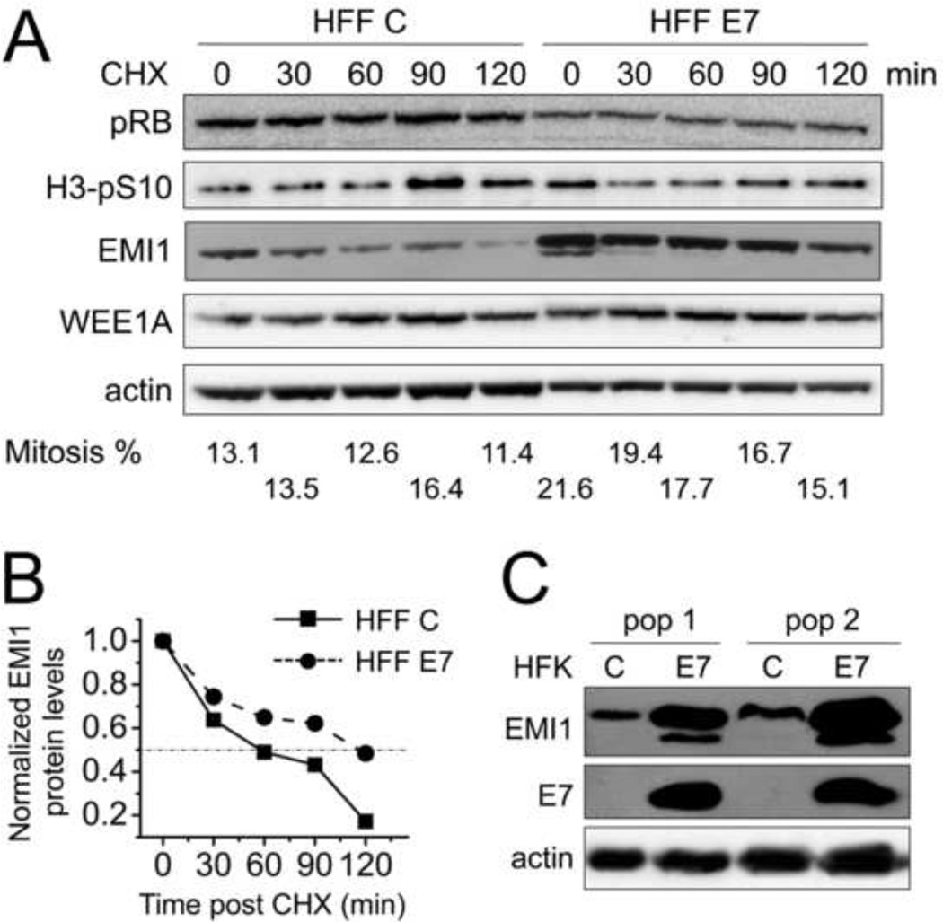
Figure 3. Mitotic EMI1 half-life is increased in HPV16 E7-expressing primary human fibroblasts.
(A) HFF C and HFF E7 were arrested in mitosis with 100 ng/ml nocodazole for 18 hours and 25 µg/ml cycloheximide was added to the medium to stop protein translation. Protein extracts were prepared at the indicated time points and steady state levels of EMI1 and WEE1A were determined by Western blotting. The levels of pRB, H3-pS10 and actin are also shown. Twice the amount lysate was analyzed for HFF C so as to render the initial EMI1 levels more comparable to those detected in HFF E7. The results of a representative experiment are shown. Similar results were obtained in two additional independent experiments. Mitotic indices, as determined by FACS analyses with a parallel set of samples stained with MPM-2 and propidium iodide, are presented below the blots. (B) Quantifications of EMI1 levels normalized to actin for the experiment presented in panel A are plotted. Protein levels in HFF C and HFF E7 populations at the 0 minute time points were each set to 1 even though they were not identical in the two cell populations. (C) Steady state levels of EMI1 as well as E7 and actin were determined by Western blotting in two independently derived human foreskin keratinocyte (HFK) populations with stable expression of HPV16 E7 (E7) or infected with control vector (C).

