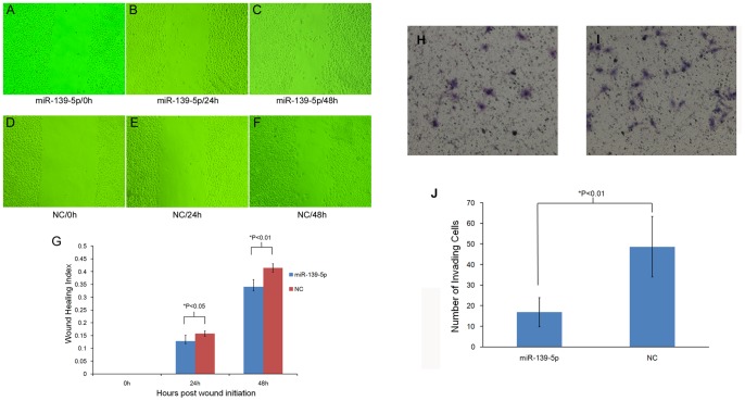
Figure 4. Cell migratory activity and invasive ability assay of EC109 cells induced by miR-139-5p.
A to F shows the effect of miR-139-5p transfection on cell migratory activity using cell scratch experiment. Wound healing was measured at 0, 24, and 48 h after wounding in miR-139-5p treated cells (A, B, and C, respectively) and negative control (D, E, and F, respectively). G represents quantitation of migration following miR-139-5p transfection versus controls. The wound healing index was defined as the ratio of (initial wound width-wound width after healing at specific time) divided by initial wound width. The result showed that miR-139-5p over-expression significantly suppressed cell migration both at 24 h and 48 h after wounding. Cell invasive ability of EC109 cells treated with miR-139-5p mimic (H) or negative control (I) was determined using a 24-well transwell plate. The invading cells transferred to the underside of the transwell membrane were stained with hematoxylin-eosin for counting. J represents the number of invading cells following miR-139-5p transfection versus controls. The results showed that the number of invading cells when treated with miR-139-5p mimic is significantly reduced compared with negative control (P<0.01).

