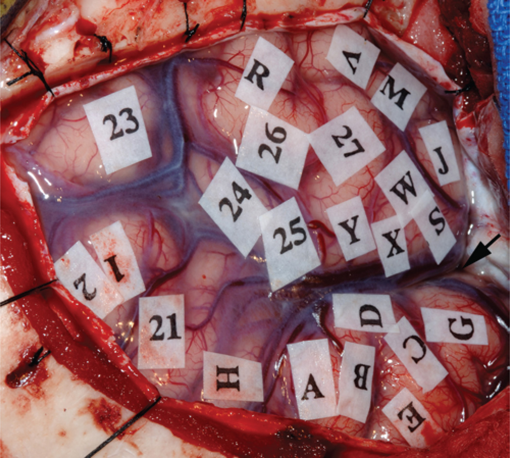
Fig. 3.
Intraoperative photograph showing the view from the surgeon’s perspective after a left frontotemporal craniotomy. The left temporal lobe is in the upper part of the photograph. The sylvian fissure is marked with an arrow. During the mapping process, sterile tags were used to mark eloquent areas of the cortex, which were associated with the following responses to stimulation: 1 and 2, paresthesias in mouth; 21, speech arrest during counting in English (Broca area); 24, errors induced during English confrontation naming test (English Wernicke area); 23–27, speech arrest during Mandarin confrontation naming test (Mandarin Wernicke area). Letters indicate regions that were not associated with any observable response to stimulation.

