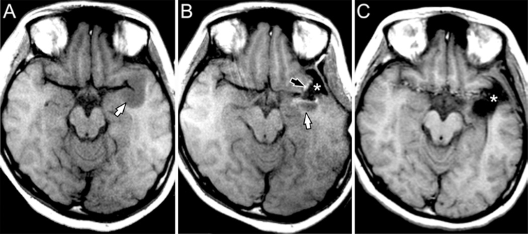
Fig. 4.
Axial T1-weighted images (from the same case as Fig. 3) demonstrating improvement in resection of nonenhancing tumor using iMR imaging guidance. A: Noncontrast image obtained in the iMR imaging scanner immediately prior to craniotomy demonstrates a hypointense mass in the left anterior temporal lobe (white arrow). B: Intraoperative MR image obtained after initial resection documents CSF signal in the resection cavity (asterisk), hyperintense signal representing blood at the margins of the cavity (black arrow), and residual tumor posterior to the resection cavity (white arrow). The operation was extended. C: Postoperative MR image shows extension of the resection cavity (asterisk) to include the region of residual tumor. No residual tumor or blood products can be identified.

