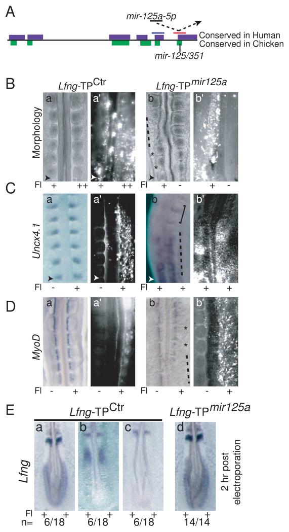
Figure 3.
Blocking interactions between mir-125a-5p and Lfng perturbs somitogenesis and segmentation clock function. A. Schematic of the Lfng 3′UTR and the TPs used, with the single mir-125a-5p binding site in chicken. Approximate positions of Lfng-TPCtrl (blue) and Lfng-TPmir- 125a (red) are shown (not to scale). B. Electroporation of Lfng-TPctr has no effect on somite morphology (panels a, a’), while electroporation of Lfng-TPmir-125a results in abnormal somite morphology with absent (dashed lines) or disorganized (*) intersomitic boundaries (panels b, b’). C. Uncx4.1 expression is disorganized and sometimes reduced in Lfng-TPmir-125a positive embryos (dashed line, panels b, b’) compared to embryos electroporated with Lfng-TPctr (panels a, a’). Note relatively normal somites in the less positive region of panel b (square bracket) D. MyoD expression in the somites of Lfng-TPmir-125a positive embryos (panels b, b’) indicates that myotomes are formed, but somite compartments are of irregular sizes (*) compared to embryos electroporated with Lfng-TPctr (panel a, a’). In some regions of the embryo, MyoD expression is strongly downregulated or delayed (dashed line, panel b). E. 2h post-electroporation, cyclic expression of endogenous Lfng is observed in Lfng-TPCtrl embryos (panels a-c), while robust, non-cyclic Lfng expression is observed in the caudal PSM of Lfng-TPmir-125a positive embryos (panel d, n=14/14). Fl: fluorescein as described in Fig. 2. Arrowheads indicate the most recent somite boundary. See Fig. S3 for analysis of target protector activity in cell lines, and Fig. S4 for fluorescein images.

