Figure 2.


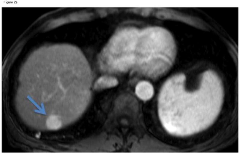
Figure 2a. T1-Weighted gadolinium-enhanced MRI reveals a focal enhancing mass in hepatic segment 7 (arrow). This mass demonstrated venous phase contrast washout, meeting the guidelines for HCC. After discussion at multidiscipline tumor board, this non-operative candidate underwent cTACE.
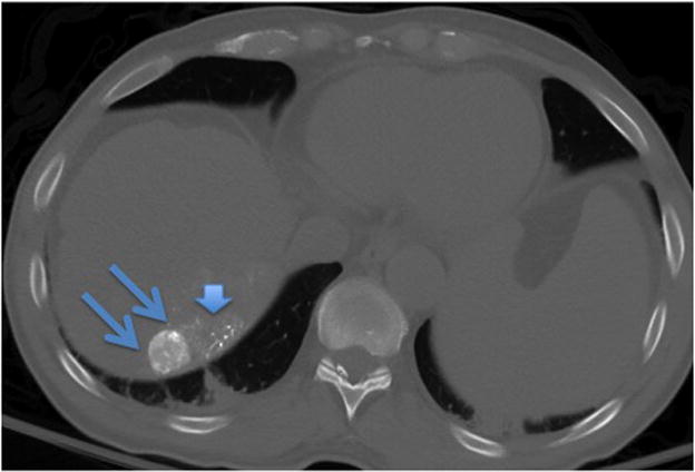
Figure 2b. A non-contrast CT scan performed immediately after cTACE revealed focal uptake of Lipiodol within the targeted tumor (double arrows). There is some non-target Lipiodol uptake in the non-cancerous hepatic parenchyma (arrow head) adjacent to the tumor.
