
Fig. 2.

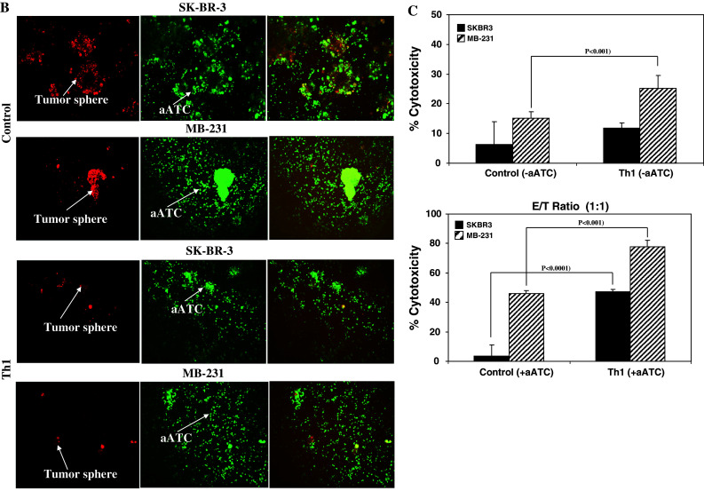
a Shows the effect of Th1 cytokines on tumor growth at day 7 using inverted microscopy. Visibly reduced size of tumor spheres for SK-BR-3 and MB-231 BrCa cells were evident under Th1 cytokines when compared with control condition (top panels, 20×; bottom panels, 100× magnification). b Confocal imaging of BrCa cells labeled with DiI (red fluorescent dye) at day 7 followed by co-culture with DiO (green fluorescent dye)-labeled aATC at a 10:1 E/T for an additional 72 h shows that armed ATC was able to engage tumor cells. In the presence of Th1 cytokines, reduced size of tumor spheres were observed (20× magnification). c Cytotoxicity quantitated by MTT assay in 2D cultures of tumor cells and aATC for 3 days at an E/T ratio of 1:1