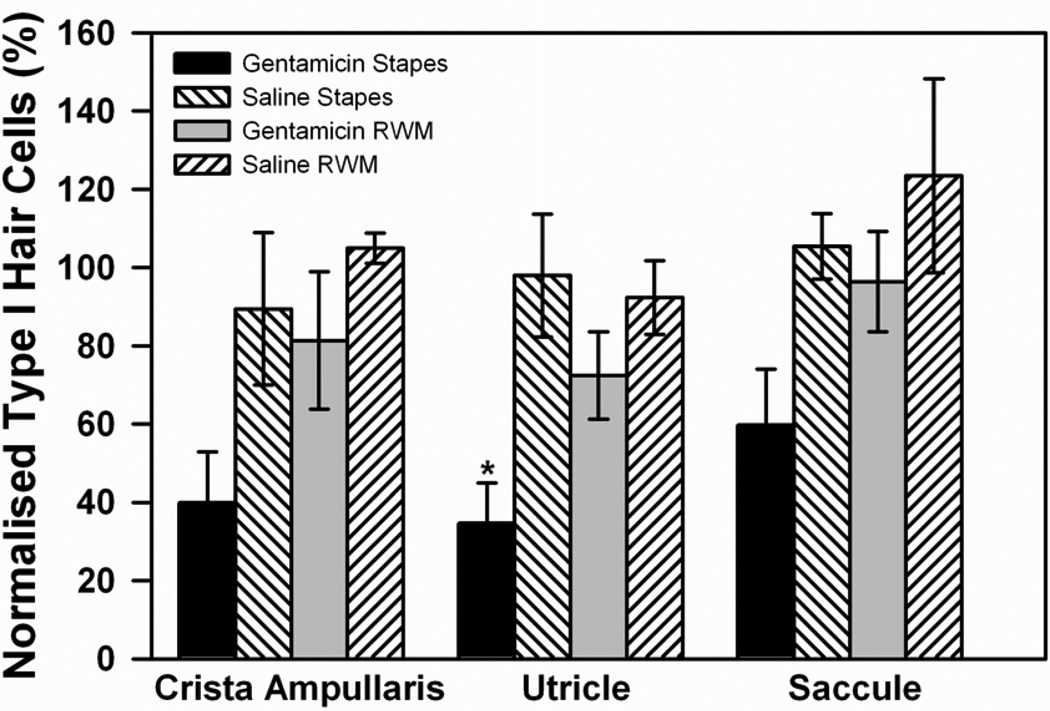
Figure 5.
Type I hair cell counts in the mid-section of crista ampullaris of the posterior semi-circular canal, the utricular macula, and the saccular macula in the vestibule. Cell counts were compared for gentamicin or saline applications to the stapes or round window membrane (RWM). In all groups, counts of morphologically normal type I hair cells were normalized with respect to those of the untreated, contralateral ear. Error bars indicate SEM.

