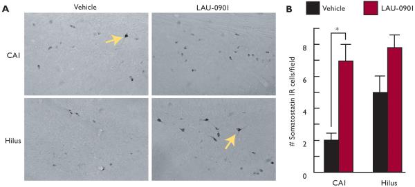
Figure 3. LAU-0901 limits somatostatin interneuronal loss in the hippocampus after status epilepticus.
(A) Representative coronal sections of CA1 and hilus showing somatostatin (SOM) IR cell profiles (arrows) from vehicle- and LAU-0901-treated rats under high magnification (40X). Note that LAU-0901 presents more IR cell profiles than vehicle. (B) Quantification of IR cell profiles showing that LAU-0901 limits SOM IR cell loss as a consequence of SE as compared to vehicle. Note that LAU-0901 significantly limits SOM cell profile loss in CA1, which reveals a trend in the hilus. Values represent averages and ± S.E.M., *=p<0.05.

