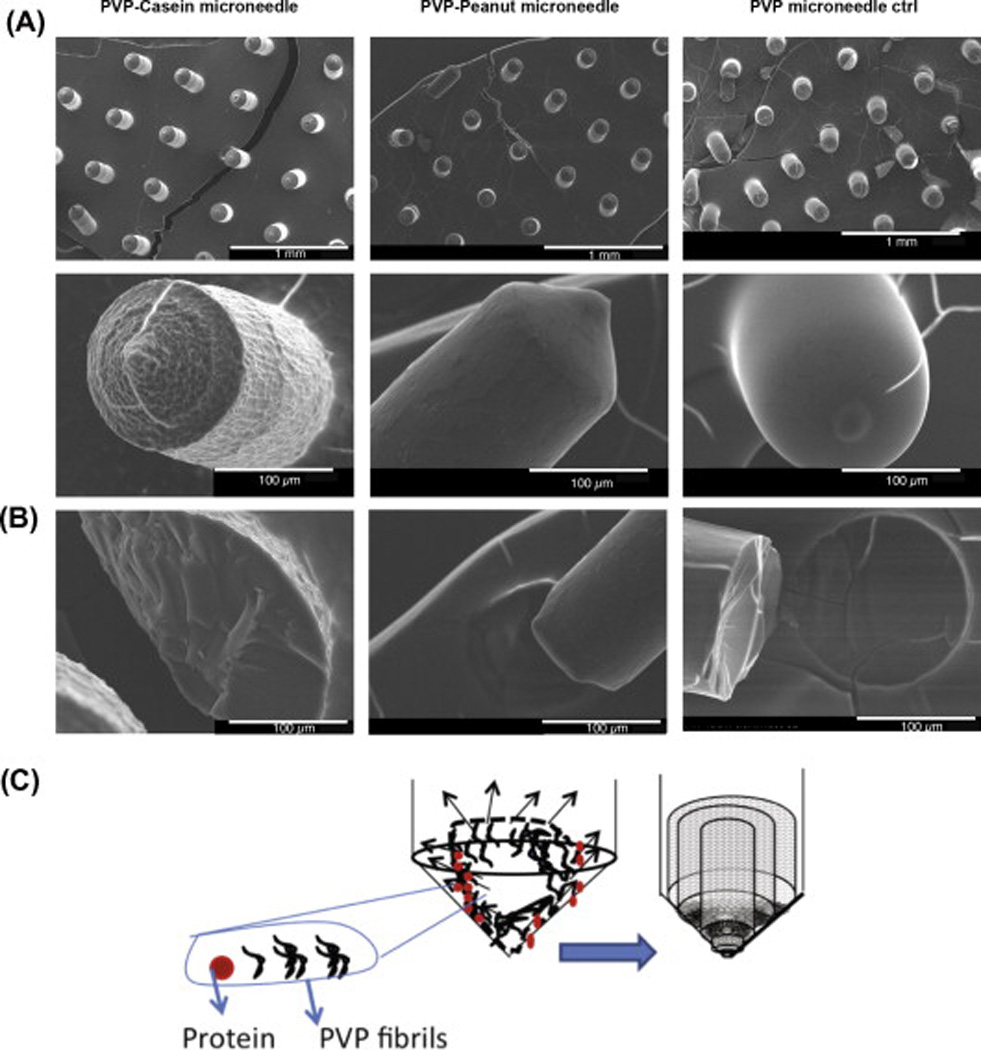
Fig. 2.

SEM images of microneedles. (A) (Row 1) Microneedle array of PVP-casein, PVP-peanut proteins and PVP alone (Control). (Row 2) Magnified single microneedle showing the surface morphology. (B) Morphology of the interior of the broken microneedle. (C) Schematic representation of the aggregate separation and packed arrangement of the PVP fibers.
