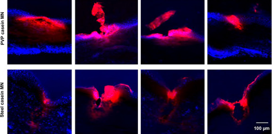
Fig. 6.

Penetration of microneedles into skin. Fluorescent images showing a comparison of the penetration of PVP-rhodamine B labeled casein microneedles and casein-coated AdminPatch array 1200 steel needles into human foreskin. Blue, DAPI staining; red, rhodamine B. Four representative penetration sites are shown for each type of microneedle.
