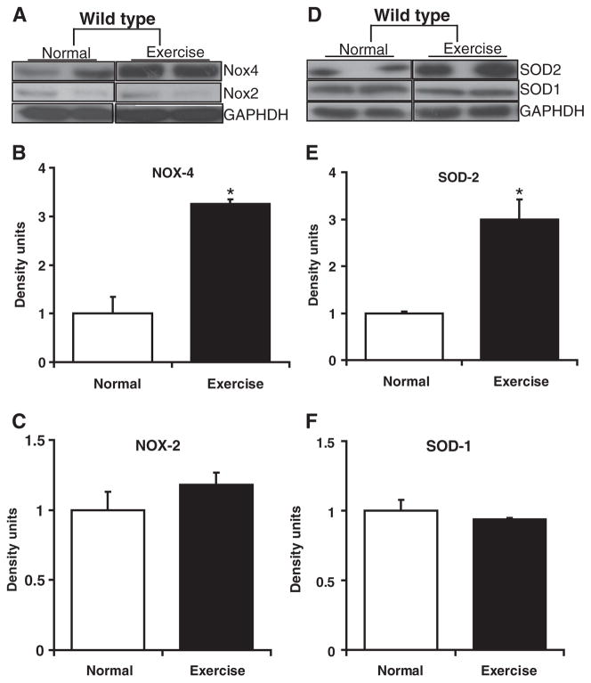
Fig. 3.
Source for reactive oxygen species (ROS) generation in the heart of AES mice. Western blots showing SOD and NOX isoforms in the sedentary and AES WT mice (n=4) myocardium. Cytosolic SOD1 (D, F) and Nox2 (A, C) levels were unaltered, while the mitochondrial Nox4 (A, B) and SOD2 (D, E) were significantly (*P<0.01) increased in AES when compared to sedentary WT myocardium.

