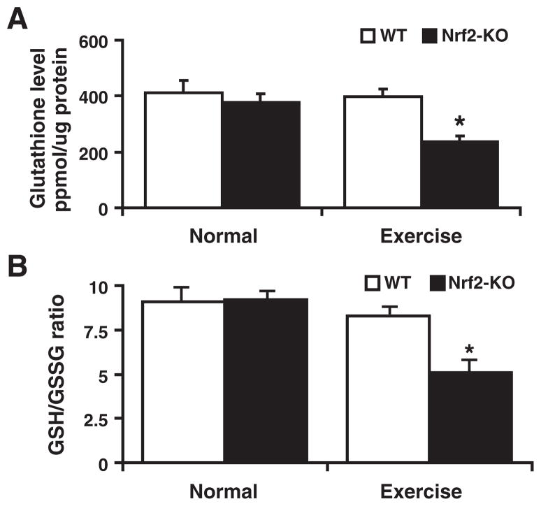
Fig. 4.
Glutathione redox (GSH/GSSG) state of the myocardium in the WT and Nrf2−/− mice: Determined the redox state of myocardial (ventricular) glutathione in WT and Nrf2−/− mice at 2 months of age (n=6) under basal and after AES conditions. (A) Under basal conditions, no statistically significant change in GSH was measured in WT and Nrf2−/− groups. Significant decrease in GSH was evident in the Nrf2−/− mice after AES, indicating onset of oxidative stress (*P<0.01). (B) Glutathione redox ratio (GSH/GSSG) was comparable under the basal state, but it was tremendously decreased in Nrf2−/− when compared to WT on AES, suggesting oxidative stress in the Nrf2−/− mice. Values are mean±SD for 6 or more animals in each group.

