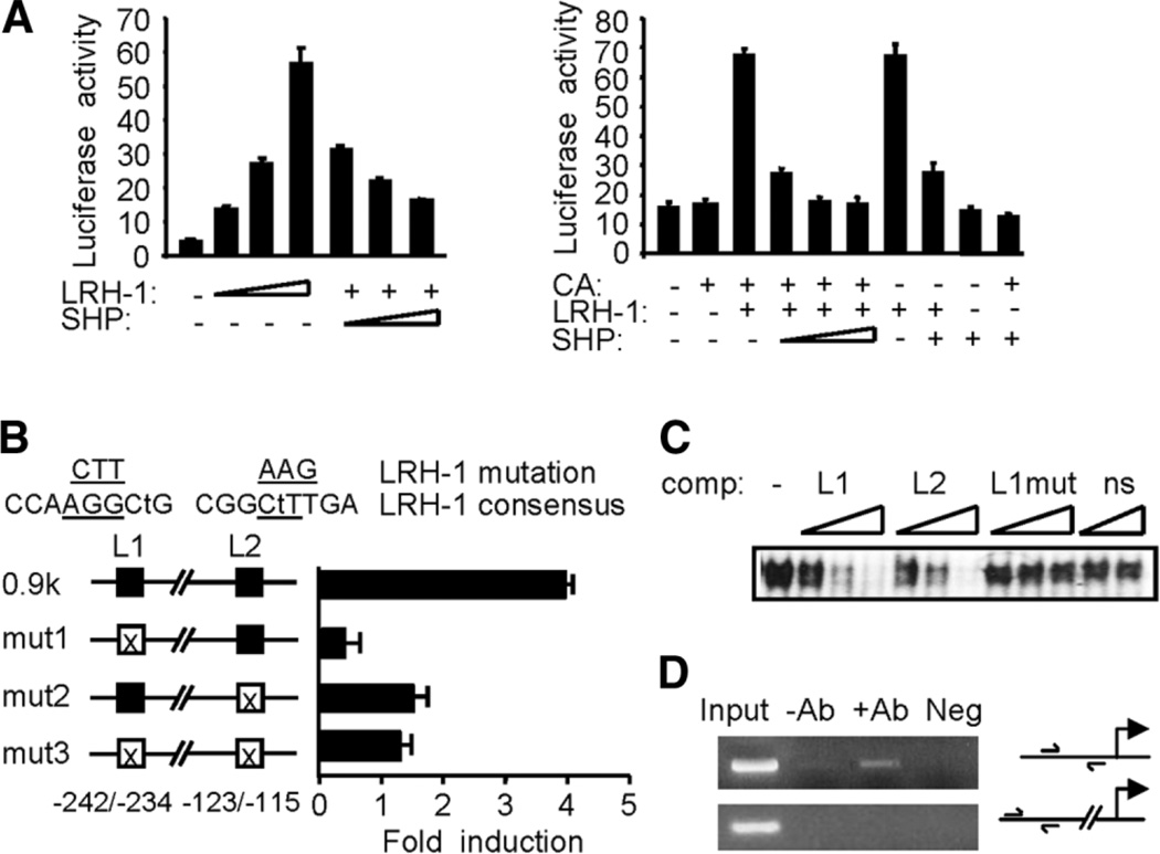
Fig. 4.
Transcriptional repression of cyclin D1 promoter by SHP. (A) Left: a − 187mCD1-Luc was transfected into CV-1 cells with LRH-1 (50, 100, 200 ng) in the absence (−) or presence (+) of SHP (100, 200, 300 ng). Right: reporter gene of −970mCD1-Luc was cotransfected with expression vectors for β-catenin (CA, 400 ng), LRH-1 (100 ng), and SHP (100, 200, 300 ng; fixed amount: 200 ng) as indicated. Luciferase activities were normalized against β-galactosidase and expressed as relative activity. (B) Mutagenesis study. The mCD1 gene contains two potential LRH-1 sites (L1, L2). Mismatches with the consensus sequence are indicated by lower case letters, and mutated sequences are underlined. The mutated constructs (mut1, mut2, and mut3) were transfected into CV-1 cells with or without LRH-1 (100 ng). (C) Gel shift assay. In vitro translated LRH-1 was incubated with the 32P-labeled L1 and increasing amounts of unlabeled L1 (lanes 2–4), L2 (lanes 5–7), and mutated L1 (lanes 8–10) or HNF-4 binding sequences (ns: lanes 11–12). (D) Chromatin immunoprecipitation analysis. Chromatin preparations from MEFs were subjected to chromatin immunoprecipitation assay with anti-SHP antibodies. DNA in the immunoprecipitates was polymerase chain reaction-amplified with the primers shown in the illustration of the promoter region. DNA from equal amounts of chromatin was polymerase chain reaction amplified before immunoprecipitation (Input). Neg, immunoglobulin G.

