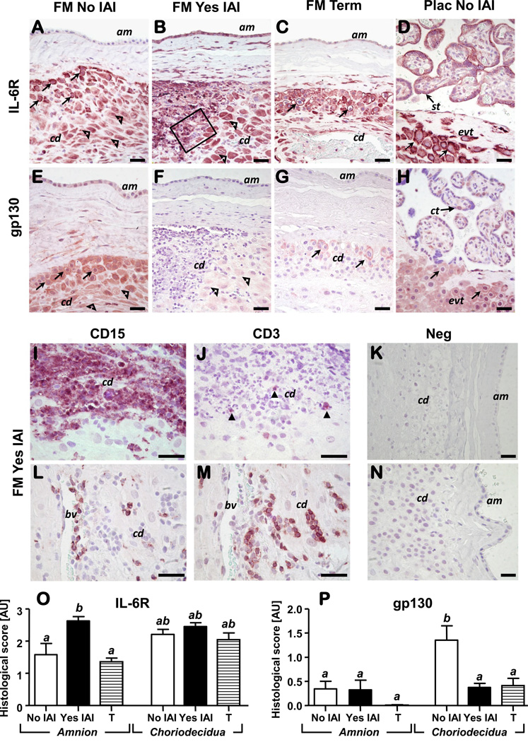
Figure 5. Representative photomicrographs of IL-6R and gp130 immunoreactivity in fetal membranes and placental sections of preterm and term pregnancies.
Preterm amnion (am) stained higher for IL-6R in the presence of IAI compared to no IAI (A-B) and term fetal membranes (C). Intense IL6-R immunostaining was identified in extravillous trophoblasts (evt, arrows) and decidual cells (open arrowheads) of the choriodecidua (cd) as identified by cytokeratin or vimentin positive staining (data not shown). In the placenta (D), IL-6R was identified in evt and villous synctiotrophoblasts (st) without notable changes with either IAI or GA (data not shown). Localization of IL-6R staining in extravillous trophoblasts (evts) was both peri-membranar and intra-cytoplasmic consistent with the ability of the antibody to recognize both membrane bound IL6-R and sIL-6R. The amnion (am) stained less positive for gp130 than choriodecidua (cd) with no discernable differences with histological inflammation or GA (E-G). Both extravillous trophoblasts (arrows) and decidual cells (open arrowheads) stained intensely for gp130 in the absence of histological inflammation (E). In the placenta (H), the strongest gp130 signal was noted in villous cytotrophoblasts (ct) and extravillous trophoblasts (evt, arrows) of chorionic plate. The outlined area in B is shown in panels I&J at higher magnification aimed to illustrate concurrent homing of neutrophils (CD15+) (I) and T lymphocytes (CD3+, closed arrowheads) (J) cells in the inflammatory infiltrate of the choriodecidua (cd) of women effected by IAI. CD15+ and CD3+ cells were also identified in the vicinity of choriodecidual (cd) blood vessels (bv) suggestive of trans-endothelial migration of inflammatory cells (L-M). Specificity of staining was confirmed by incubation of slides with rabbit IgG as control for IL- 6R & CD3 staining (K). Mouse non-immune IgG served as negative control for gp130 and CD15 mono-clonal antibodies (N). IL-6R and gp130 histological scores in the amnion and choriodecidua of preterm (NO & YES IAI) and term patients (T) is shown in panel O and P, respectively. Data presented as mean + SEM and analyzed by one-way ANOVA followed by post-hoc Student-Newman-Keuls tests. Means with at least one common superscript are not statistically significant (P > 0.05). Scale bars: 30 µm for all panels (A-N).

